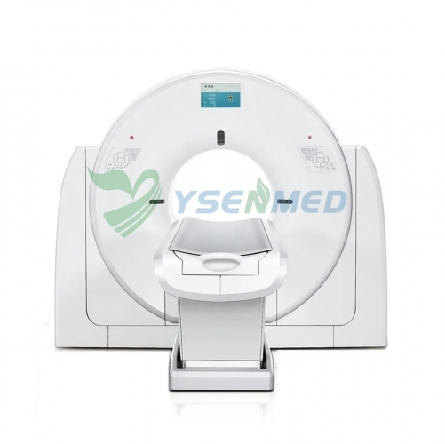
Scanner CT double énergie 128 coupes YSCT-128C
| Product parameters | |
| Brand: | YSENMED |
| MOQ: | 1 |
| Package: | Wooden Package |
| Ship From: | Guangzhou |
| Lead Time: | 7 days |
| Customization: | Customized packaging |
Description
Computed Tomography

Advantages:
a)Precise P-Axial tomography imaging technique;
b)Multi-pixel HD algorithm;
c)Dual-energy platform;
d)Low dose control system;
e)Intelligent post-processing function;
2.Characters
1)Precise P-Axial tomography imaging technique
P-Axial tomography imaging technique is the patented technique, which offers the minimum slices thickness of 0.312mm ultra-thin image. Microstructures are able to be seen, such as inner ear, pulmonary nodules.
Apply to:
Microstructures of inner ear: Cochlear, bony labyrinth, membranous labyrinth, ear bones, ligaments and nerves of ear bones all can be seen clearly, it will make the diagnosis of inner ear more accurate, especially qualitative measurement
Pulmonary nodules: The clear structure and character of early pulmonary nodules avoid the wrong diagnosis and overtreatment of pulmonary nodules
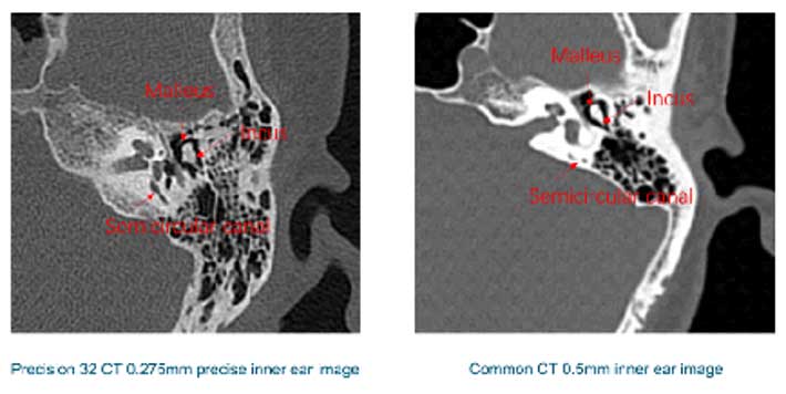
YSCT-128C 0.312mm precise inner ear image
Common CT 0.5mm inner ear image
High quality images are closely related to advanced CT reconstruction algorithm, we independently develop the Hi-Resolution HD algorithm, combine with 1024x1024m image matrix, the human body and lesion structure could be displayed to the extreme.
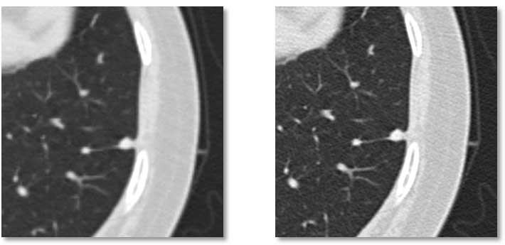
Lung image by normal algorithm
Image by multi-pixel HD algorithm
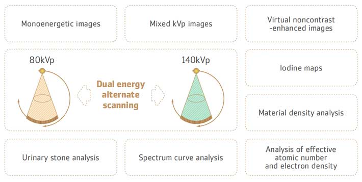
a)Homology: In the collection mode of homology of an X-ray tube, the interference of scattering line artifacts between two tubes is perfectly avoided.
b)Homodomain: The bed is stationary during the X-ray data collection of high and low kV energy, ensure accurate matching of high and low kV data positions and reducing image artifacts caused by position mismatch.
c)Simultaneous: The 0.72 second dual-energy scan can achieve a wide range of dual-energy images very fast, and realize the collection of simultaneous image of whole organ.
In addition, YSCT-128C provides ultra-low-dose dual-energy imaging technique with a minimum of 60 mA, combine with dual-energy noise reduction technique, to make sure the radiation dose of dual-energy scan lower than normal scan, while obtain excellent images. In addition, there will be no tube overheating protection due to kV fast switching.
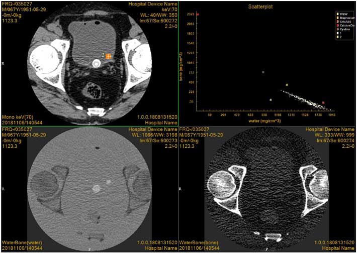
Case presentation: This patient was a 67-year-old man with many years of bladder calculi. Image showed two large (1) small (2) stones in the bladder, and the main components of the stones were determined as uric acid by scatter plot. At the same time, calculi were found in the water diagram but not in the bone diagram, which was also consistent with the characteristics of uric acid calculi.
In order to minimize the radiation dose to patients, YSCT-128 includes the low dose control system such as P-Dose, P-IR and so on, also provides scanning solution specialized for child, combine with 60kV ultra-low-energy CT scans and accurate dose modulation of P-Dose, as well as parameters optimized at the appropriate dose level specifically designed for child care, it can reduce 80% radiation dose. P-Dose can intelligently adjust the scanning dose of different parts of human body according to the density and shape of different parts of human body. Advanced P-IR iterative reconstruction algorithm guarantees the high quality image in each low-dose scanning mode.
60kV Ultra-low-dose CT Scanning:60kV/70kV ultra-low energy X-ray CT scan mode which is the lowest in the industry, reduce the damage of CT radiation to the patients by decrease of the X-rays energy.
P-Dose 3D Precise Milliampere Modulation: Depending on the density and shape of the patient’s scanning site, the exposure dose of each View can be adjusted accurately, in addition to intelligently adjusting the scanning dose of each site.
P-IR Advanced Dual-domain Iterative Reconstruction: Multiple iterations of noise reduction in the original and image data Fields, enhance image quality
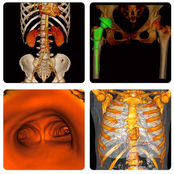
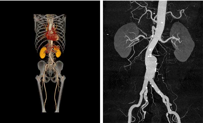
5)Intelligent post-processing function
The post-processing workstation of YSCT-128C, follow DICOM3.0 international standard, realizes to obtain, process, analyze, manage, archive, query, browse, print, standard image format output of DICOM, report writing and so on.
VR、MPR、CPR、MIP、AIP、MinIP、SSD and other image;
Automatic bone removal, bed plate removal;
Virtual endoscope;